
Equipment
Our microscopes include widefield, TIRF, super-resolution, lightsheet and point scanning confocal with nonlinear optics instruments. The setups are equipped for live-cell imaging, FRET, Ca2+ imaging, slide scanning, label-free imaging, FRAP/photo-activation and more. We can accommodate a range of samples, from single molecules over cells and tissue sections to centimeter large specimens.
Microscopes
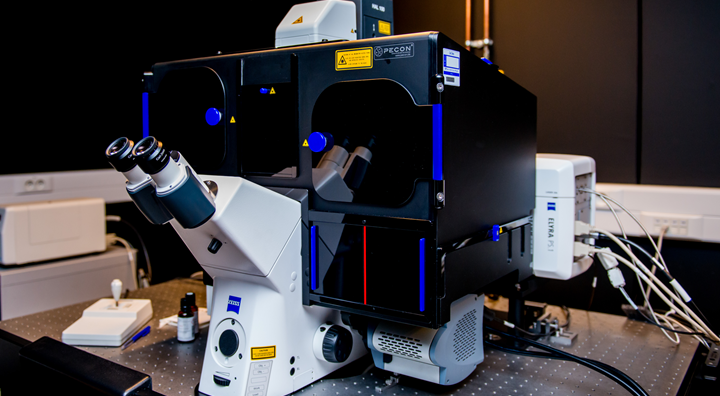
Zeiss Elyra PS.1
Perform fast, live-cell fluorescence imaging experiments on this powerful widefield fluorescence microscope with TIRF and 3D super-resolution (SIM and SMLM) capabilities. This commercial Zeiss microscope is equipped with 4 laser lines (405, 488, 561 & 642nm), an incubation chamber, a dedicated SIM camera and SMLM camera, Definite Focus system and piezo stage. Additionally, the microscope is combined with a patch clamp setup allowing simultaneous fluorescence and electrophysiology recordings.
Widefield - TIRF - super-resolution
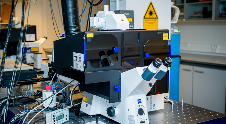
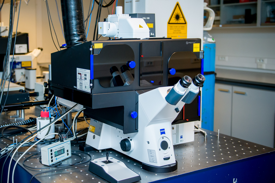
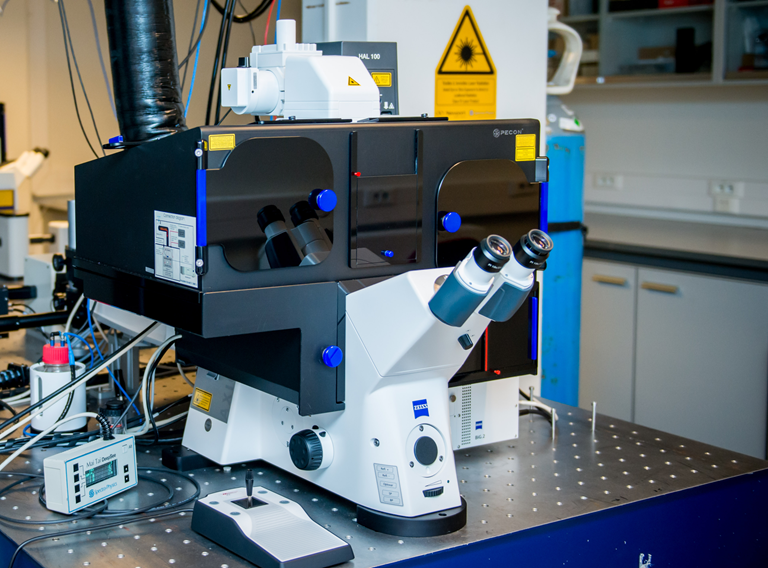
Zeiss LSM880 NLO AiryScan
Use this highly versatile state-of-the-art confocal microscope to solve all your multi-dimensional research questions. Equipped with an NLO module for deep-tissue and label-free imaging, pulsed excitation and time-resolved detection for FLIM, a spectral detector, and an AiryScan detection system for super-resolved fluorescence microscopy, the LSM880 provides a convenient solution for most of your imaging needs.
Confocal - two-photon - label-free - correlation - super-resolution - spectral detection

Zeiss LSM900 Airyscan 2
Use this highly versatile state-of-the-art confocal microscope to solve all your multi-dimensional research questions. Equipped with sensitive GaAsP PMT detectors, flexible variable beam splitter dichroics and the latest Airyscan 2 detection system for super-resolved fluorescence microscopy, the LSM900 provides a convenient solution for most of your imaging needs. Additionally, the AI Sample Finder and advanced Navigation and Tiles module make it very easy to find regions of interest and set up your imaging experiments.
Confocal - multidimensional - correlation - super-resolution
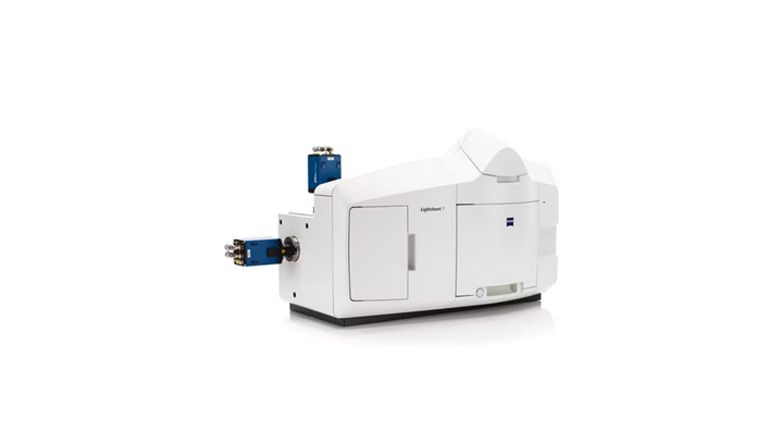
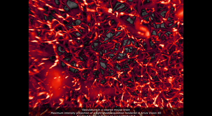
Zeiss Lightsheet 7
Use the Lightsheet 7 for fast volumetric imaging of sensitive samples such as living model organisms and organoids, or image large cleared samples as a whole with sub-cellular details. The fully incubated microscope has 5 laser lines spanning the visible spectrum and is equipped with two cameras for simultaneous dual-color acquisitions. A broad range of objective lenses and sample chambers can accommodate almost any sample.
Light sheet - volumetric (3D) imaging - gentle illumination
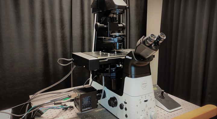
Nikon Eclipse Ti2-E
This fully motorized microscope with an exceptional field-of-view is equipped with a powerful multicolor LED lightsource and a Photometrics Kinetix camera for very fast imaging of large sample areas, making it the perfect match for all your functional imaging needs.
Widefield - LED excitation - fast - big FOV
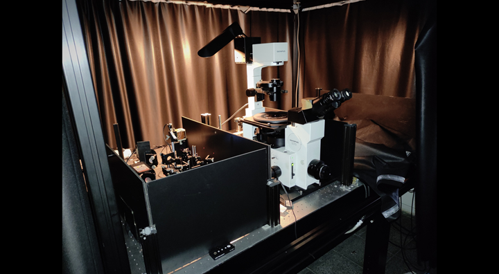
Homebuilt smFRET
Homebuilt dedicated 483/635-nm alternating excitation, dual-color dual-polarization detection confocal microscope. This system allows imaging more than 20 parameters per single molecule, making it the ideal system for subnanometer accurate single-molecule FRET studies.
Pulsed Interleaved Excitation (PIE) - single-molecule FRET - single-molecule fluorescence - FCS
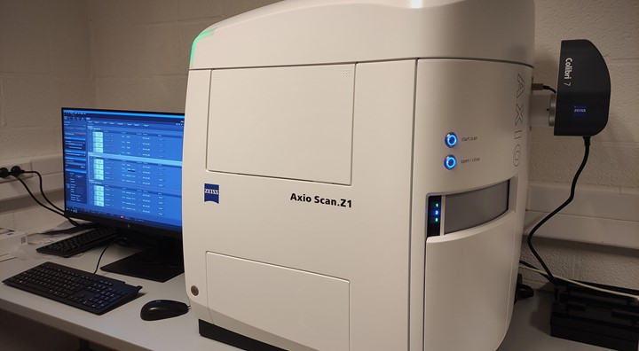
Zeiss Axioscan Z.1
Increase the throughput of your experimental workflow with this fully automated whole slide scanner. Equipped with a multicolor LED lightsource, high resolution lenses, automated focussing and positioning control, and high quality detection optics, the upright microscope captures brightfield and fluorescence images of outstanding quality. The ZEN software suite allows simple and automated digitization of your (large) samples.
Slidescanner - automation - widefield - brightfield - fluorescence
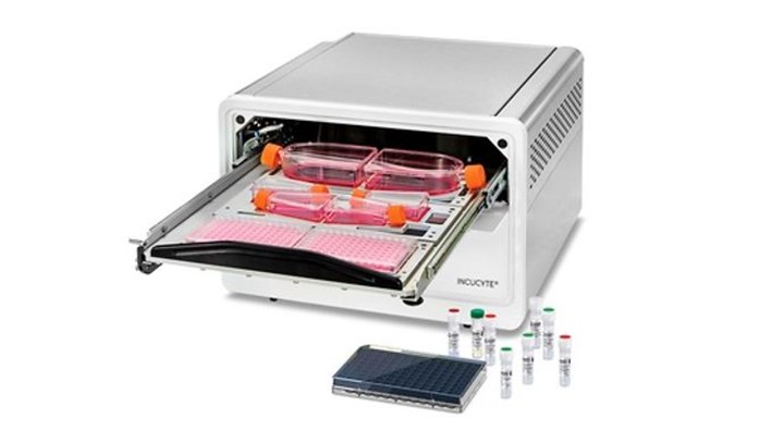 IncuCyte S3 Life cell imager
 IncuCyte S3
Use the Live-Cell Imaging and Analysis System to automatically acquire and analyse cells in a physiological environment. Analyze even the most sensitive living cells around the clock for days, weeks or months and investigate cell health, perform functional assays, or discover morphological and phenotypic changes.
Life cell imager - automation - physiological conditions
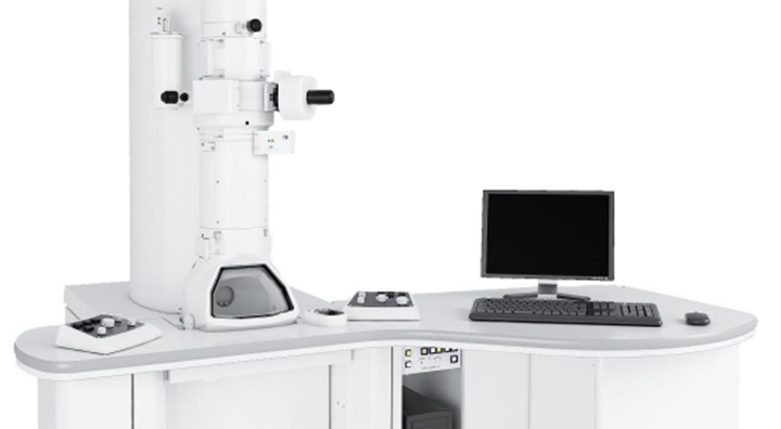 Jeol JEM-1400Flash
 Jeol JEM-1400Flash
Acquire ultrahigh resolution images of your biological samples or nanotechnology, polymer, and advanced materials. This compact TEM combines excellent resolution and ease of use. Couple functional fluorescence imaging with ultrastructural TEM information in Correlative Light Electron Microscopy (CLEM) to gain in-depth knowledge on a subcellular and molecular level
Transmission Electron Microscopy - ultrastructure - Correlative Light Electron Microscopy
Processing and analysis
Computing Capacity
Speed up your data processing and analysis procedures using the dedicated analysis workstation.