
Oligodendrocyte biology and remyelination platform
- Are you developing new therapies for neurodegenerative disease that target oligodendrocyte biology or de- and remyelination?
- Progress your development programs faster by building on our expertise glial cell biology, neuroscience, and lipid metabolism.
- Our validated models are continuously exploited in proprietary research programs and service activities for the private sector.
- We are keen on creating collaborative and longlasting partnerships by meeting rapidly changing needs and requirements in a flexible way and delivering on our promises.


POSSIBLE APPLICATIONS
- Test the therapeutic activity of candidate therapies on oligodendrocyte biology and remyelination in vitro at three levels: differentiation and proliferation of oligodendrocyte precursor cells (OPCs), migration behaviour of OPCs and axon-wrapping potential. Multiple conditions can be tested in parallel.
- Test the effect of therapies in a more complex multicellular brain environment using ex vivo in brain slice cultures. Multiple conditions can be tested in parallel.
- Test your candidate therapies in in vivo models which includes a battery of functional, structural, and clinical outcome measures.
IN VITRO MODELS
DIFFERENTIATION OF MOUSE OPCS
Read-outs: Expression of myelin proteins (PLP/MBP), number of OPCs and oligodendrocytes, process complexity immunostaining, gene expression (qPCR)
(example published as Dierckx et al. 2022; 119: e2120393119)
Phloretin stimulates oligodendrocyte precursor cell maturation in vitro. Representative immunofluorescence images of OPCs treated with vehicle or phloretin and stained for O4 and MBP.
Scale bar, 25 μm.

MICRO-FIBER MYELINATION ASSAY
Read-outs: Immunocytochemistry for MBP Imaging with confocal imaging on mouse OPCs.
(example published as Schepers et al. Brain Behav Immun 2022; 109: 1-22)
Confocal and 3D rendered images showing the formation of myelin-like extensions on microfibers by primary mouse OPCs.

AGAROSE DROP MIGRATION ASSAYS
Read-outs: cell tracking using Incucyte
Agarose is added to cell suspension and dropped at the center of wells in a 24-well tissue culture plate. The extent of cell migration is measured after 5 days using Incucyte imaging analysis.
(see publication Willems et al. FASEB J. 2024 Jan 31;38(2):e23413. doi: 10.1096/fj.202301557RR)

EX VIVO MODEL
CEREBELLAR BRAIN SLICE MODEL
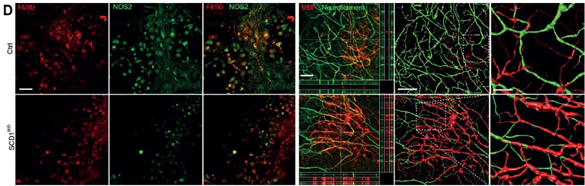
Read-outs: Microglial lipid load (ORO), myelination of axons (MBP/neurofilament immunostaining), inflammatory mediator expression (qPCR)
(example published as Bogie et al. J Exp Med. 2020; 217: e20191660)
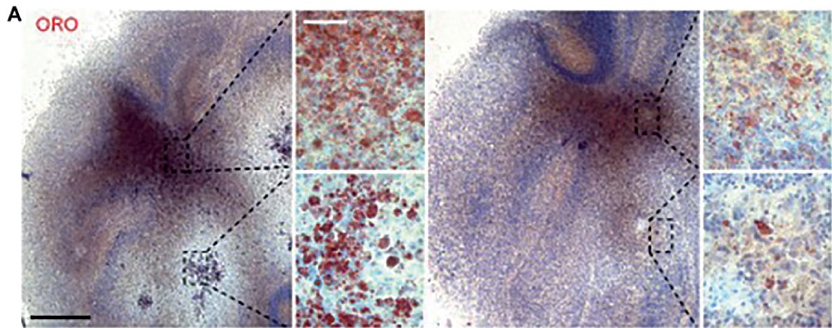
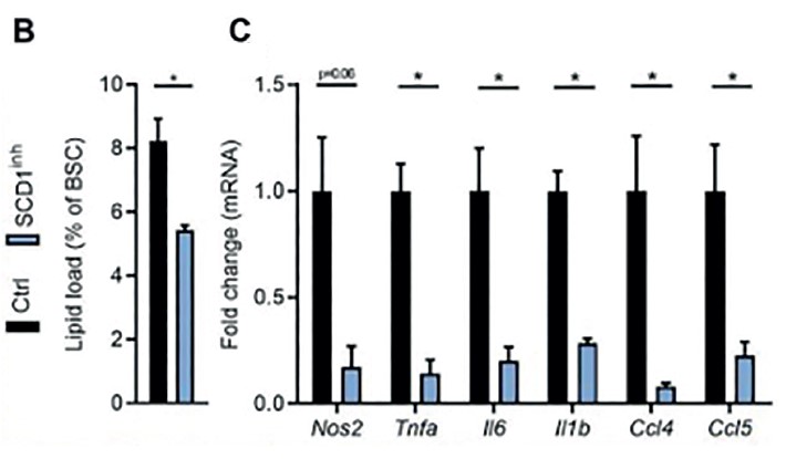
(A and B) Representative images and quantification (lipid load defined as percent area covered in lipid droplets of the total brain slice area) of ORO (EC) staining of cerebellar brain slices treated with an SCD1 inhibitor or vehicle (n = 3 slices). Scale bars, 500 μm (overview); 50 μm (inset). (C) mRNA expression of inflammatory mediators in cerebellar brain slice cultures treated with an SCD1 inhibitor or vehicle (n = 4 slices). (D) Representative immunofluorescence images of brain slice cultures treated with vehicle or an SCD1 inhibitor and stained for NOS2/F4/80+ (n = 3 slices; scale bar, 50 μm) and MBP/neurofilament (n = 3 slices; scale bar, 50 μm; orthogonal and three-dimensional reconstruction).
ANIMAL MODELS TO STUDY DE/REMYELINATION
CUPRIZONE MODEL: ACUTE OR CHRONIC DEMYELINATION
Molecular read-outs: Myelin visualisation (Luxol Fast Bleu staining), myelin layer thickness (TEM G-ratio), gene expression (qPCR), protein expression of myelin protein (MBP)
(example published as Houben et al. PNAS 2020; 117:5028-5038)
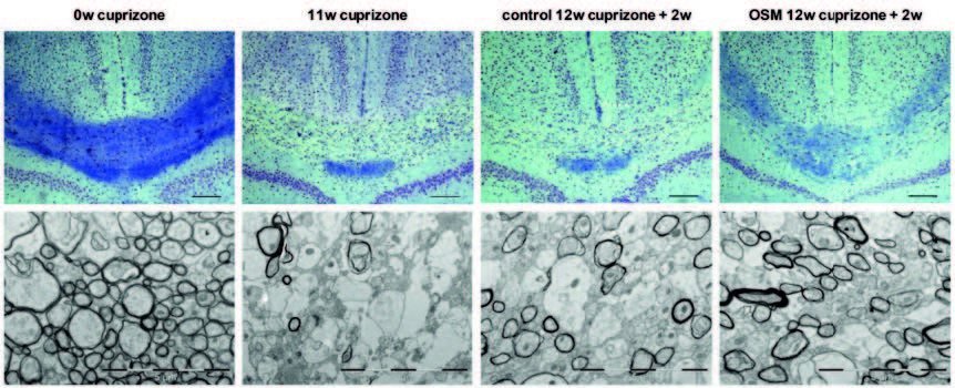
Representative images of LFB staining in the corpus callosum (around bregma −1.82 mm).
Representative TEM images and quantification of the G ratio in the corpus callosum)
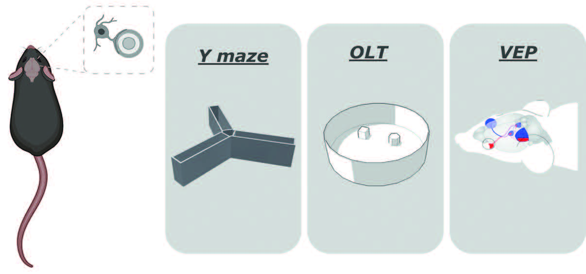
Functional read-outs: spatial memory (Y-maze and object location task), visual evoked potentials.
(see publication Willems et al. FASEB J. 2024 Jan 31;38(2):e23413. doi: 10.1096/fj.202301557RR)
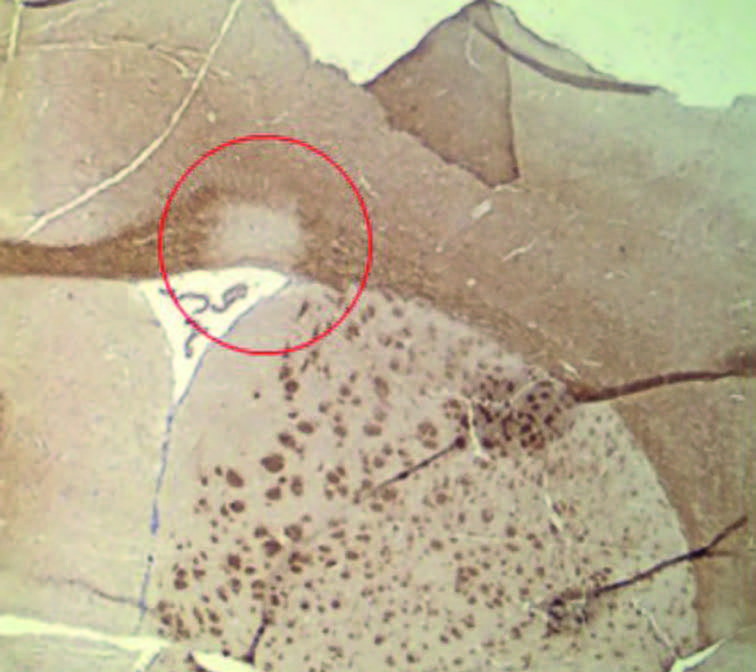
LYSOLECITHIN MODEL WITH FOCAL INJECTION
Molecular read-outs: Myelin visualisation, gene expression (qPCR)
Representative images oLPC (L-α-Lysophosphatidylcholine) is injected locally into the brain of the mouse, inducing demyelinating lesions in a specific region. The resulting dysfunction and remyelination can be subsequently assessed and analysed. Depending on the injection site, de- and remyelination can be studied in different anatomical regions of the mouse CNS.
The red circle in the figure displays corpus callosum demyelination, 7 days post injection at a time point that the myelin debris has been cleared by the phagocytes and remyelination is not yet visible. The timing after the injection determines whether phagocytes are present in the lesion (demyelination) or whether OPCs are infiltrating to study remyelination.
CHRONIC EXPERIMENTAL AUTOIMMUNE ENCEPHALOMYELITIS
Molecular read-outs: myelin layer thickness (TEM G-ratio), expression of myelin protein (MBP), gene expression (qPCR)
Functional read-out: neurological score, spatial memory
(see example published as Schepers et al. Brain Behav Immun 2022; 109: 1-22)
COLLABORATION OPTIONS
- Fee-for-Service: performing the relevant experiments for you
- Consultancy: guiding your experimental set-up
- Research collaboration: open for joint grant applications when the project is complementary with our own research lines and goals
PUBLICATIONS
BUSINESS MANAGER
Dr. An Voets

Would you like this information in a pdf? Click here.