Advanced Microscopy @UHasselt
- Development of cutting-edge microscopy
- Application of imaging in the life/material sciences
- Imaging consultancy and services


POSSIBLE APPLICATIONS
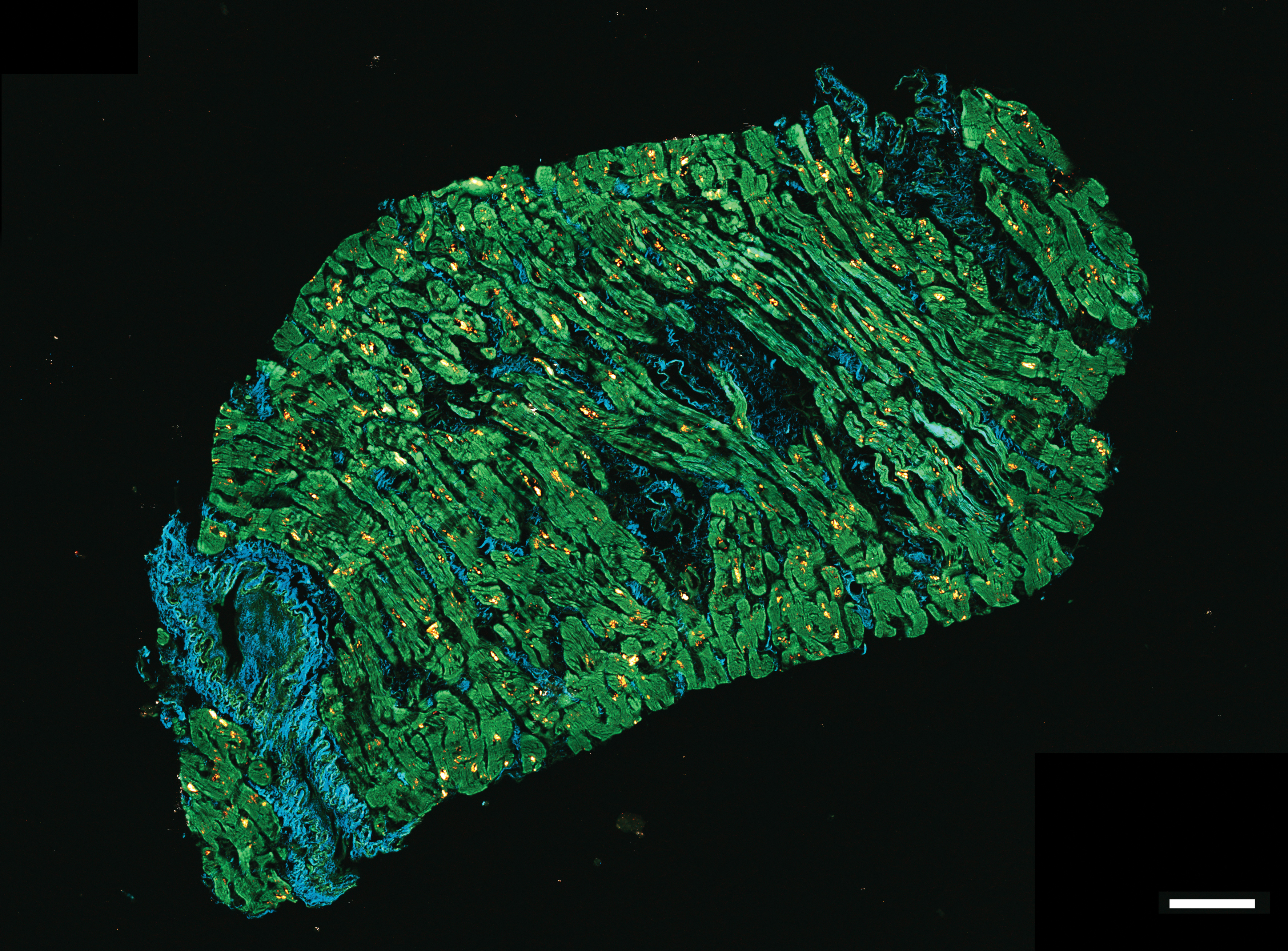
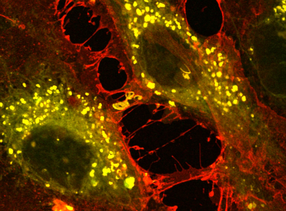
- Visualize processes and structures on a multicellular to subcellular scale in living and fixed samples.
[Brightfield, fluorescence, confocal, slidescanner, TEM, Incucyte] - Investigate molecular interactions and dynamics with high spatio-temporal resolution.
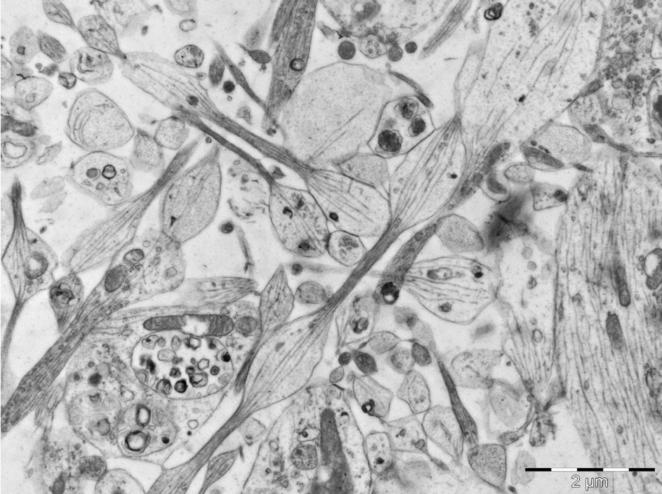
[FRET, correlation spectroscopy methods, single-particle tracking] - Zoom in on the nanometer scale using super-resolution light microscopy and transmission electron microscopy
[Airyscan, SIM, SMLM (dSTORM, PALM, PAINT), SOFI, TEM] - Apply non-linear imaging methods for deeper penetration and label-free imaging.
[Two-photon excitation microscopy, SHG imaging, White Light generation] - Probe cellular activity by combining fluorescence imaging with patch clamping.
[patch clamp fluorometry] - Study long-term cell proliferation, migration, or survival in a controlled environment
[Incucyte]
RELATED AVAILABLE SERVICES
- Cell culture, incubation, fixation
- Histological preparation of samples
» Paraffin embedding and sectioning of soft and decalcified tissues
» Resin embedding, ultrathin sectioning and tissue contrast staining
» Critical point drying for SEM specimens
» Bone microtomy
» Standard Histological stainings: Haematoxylin-Eosin, Masson’s Trichome, Alizarin red S, Alcian Blue, Cresyl Violet, Oil red O, …
» Immunohistochemistry/-cytochemistry and immunofluorescence
EQUIPMENT
Confocal set-ups
- Zeiss LSM880-Airyscan-NLO
- Zeiss LSM510-META-NLO
- SpectraPhysics MaiTai DeepSee 100 fs pulsed titanium sapphire 690-1050 nm
Widefield set-ups
- Zeiss Elyra PS.1 (widefield, TIRF and super-resolution)
- Nikon Ti2-E (ultrafast widefield with large field-of-view)
- Leica stereo microscope M60 with camera IC80HD (animal facility)
Bright field and fluorescence microscopy
- Leica DM4000LED (with color camera DFC450C)
- Leica face-to-face microscope (multi viewer DM2000 LED with camera)
Slide scanner
Zeiss Axioscan Z1(automatic slidescanner, both brightfield and fluorescence)
Incucyte Live cell analysis system
Multiplex assays in 6/24/48 and 96 well plate format; 4x, 10x, 20x objectives, phase-contrast and fluorescence images (in green and red channel)
Electron microscopy
JEOL JEM-1400Flash 120 kV Transmission Electron Microscope (+ Correlative light-electron microscopy)
EM sample prep (Cryostats and microtomes)
- Cryostat Leica 3050
- Cryostat Bright OFT-5000
- Microtome Leica Histocore Biocut
- Sample preparation: Leica EM ICE - high pressure freezer
- Cryosubstitution: Leica EM AFS2 - Automatic Freeze Substitution System
- Ultramicrotome Leica UC6
- Ultrastainer Leica
More details: www.uhasselt.be/aomc
COLLABORATION OPTIONS
- Fee-for-Service: performing the relevant experiments for you
- Facility access: once trained, you can perform experiments independently
- Consultancy and training: guiding your experimental set-up and training researchers at your location or at our facilities
- Research collaboration: open for joint grant applications when the project is complementary with our own research lines and goals
