Muscle, organ and cell physiology
Skeletal muscle physiology, organ communication & integrative exercise physiology and low back pain.
View the content of this page
Physiology of Skeletal Muscles
Our muscles are constantly adapting in response to training, aging or disease. These changes can be observed in muscle strength, endurance, or the ease with which we perform daily activities. However, these visible effects result from countless cellular and molecular adaptations within the muscle tissue. At REVAL, we therefore study skeletal muscles from the cellular level to everyday muscle function. Using this approach, we aim to understand how muscle function declines due to disease, injury or aging, and how it can be improved through exercise, rehabilitation or targeted lifestyle interventions.
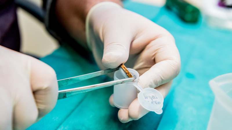
To achieve this, we combine a wide range of complementary research techniques. We measure muscle volume and strength across different muscle groups and link these functional measurements to mechanistic analyses. For example, we use near-infrared spectroscopy (NIRS) to assess muscle oxygen consumption. In addition, we collect muscle biopsies from both healthy volunteers and patients, which we then analyze at structural, molecular, and functional levels. This allows us, for instance, to determine whether changes in muscle function are driven by differences in the force that individual muscle fibers can generate or by specific molecular adaptations.
Through this integrated approach, we bridge what happens inside our muscle cells and how muscles actually perform in daily life. Our analyses are carried out in a wide range of populations, including people with obesity, COPD, low back pain, or cancer, as well as older adults and athletes recovering from injury.
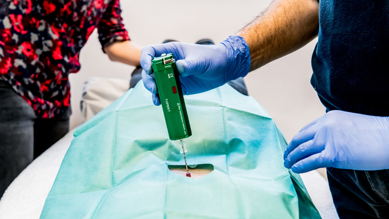
Within the REVAL research group, Prof. Dr. Frank Vandenabeele and his team have developed extensive expertise in studying the microscopic characteristics of skeletal muscle fibers (using immunofluorescence) in muscle samples obtained from healthy subjects and various patient populations (MS, DM, obesity, COPD, low back pain, COVID, cancer). These structural muscle properties are studied in relation to the functional properties of these muscles and muscle volume (determined by 3D free-hand ultrasound). Muscle samples are obtained using a "fine needle" biopsy technique, guided by ultrasound.
Chiel Poffé

Organ crosstalk and integrative exercise physiology
Each individual is made up of trillions of cells, which communicate with their environment but also internally to enable specific tasks and collaboration between cells. The cooperation between cells within a certain tissue, and even between cells in different tissues, is key for our bodily functions. This crosstalk is organized in many ways, hormones being the best known example. It has recently been shown that cells also communicate via the secretion of small bubble-like structures, called extracellular vesicles, which can be released by all body cells. The content of these cellular vesicles more or less reflect the state of the sender (cell of origin) and the processes that happening within this cell. The extracellular vesicles, and in particular their content, may in turn influence the behaviour and/or metabolism of the recipient cells.
During physical exercise, these extracellular vesicles are also prominent (e.g. in the bloodstream). Skeletal muscle is considered an important source of circulating extracellular vesicles during exercise. To date, the relative contribution of different metabolically important tissues to the circulating extracellular vesicles and their destination is largely unknown. Research on the role of exercise and/or exercise training and their relation to extracellular vesicles is still in its infancy, a field in which we have recently focused on in our research group.
Kenneth Verboven
Dominique Hansen
Low back pain
Back To Back: the forgotten role of back muscle characteristics to tailor exercise therapy for recurrent non-specific low back pain
Non-specific low back pain (NSLBP) is the leading cause of disability worldwide. During the past decade, research mainly focused on psychosocial causes of NSLBP (e.g., fear of movement), thereby reducing interest in biological causes (e.g., back muscle dysfunction).
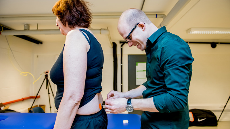
New insights into the characteristics of back muscles are, however, crucial. Our research group can provide this through our unique complementary expertise with state-of-the-art methods and our pilot results. We hypothesize that patients with NSLBP show alterations in the back muscles at macroscopic (i.e., volume, fat fraction), microscopic (i.e., fiber types), hemodynamic (i.e., oxygenation), and electrophysiological (i.e., activation) level, which are interrelated and underlie impaired back muscle proprioception in this population. This will be examined by three objectives:
- To assess the reliability and validity of 3D freehand ultrasound to measure macroscopic characteristics of the back muscles,
- To assess macroscopic, microscopic, hemodynamic as well as electrophysiological back muscle characteristics in patients with NSLBP and healthy controls, their interrelatedness, and their correlation with back muscle proprioception, to delineate phenotypes,
- To assess, by proof-of-concept, the reversibility of different back muscle characteristics by tailored exercise therapy in patients with NSLBP.
This study is funded by FWO (G072122N).
Lotte Janssens