Research
The medical dosimetry team performs research in fields such as dosimetry audits, dosimetry for biological experiments, microdosimetry, nanoparticles, small animal irradiations and dosimetry in small fields and kV X-rays.


View the content of this page
Dosimetry in kV X-rays and small fields
Introduction
Lots of improvements are happening recently in the preclinical research for radiotherapy where they are working with small animal irradiators. These machines use medium and lower kV X-rays and small fields. Besides, electronic brachytherapy sources using miniature X-ray sources are also gaining interest. To use alanine/EPR dosimetry in these conditions, its response must be characterised. The goal of this work is to investigate the relative response of alanine/EPR dosimetry at 50 – 225 kV X-rays and small fields.
Materials and methods
Alanine and film dosimeters were irradiated on two different small animal irradiators: the SARRP and the X-RAD 225CX. The detectors were irradiated with beams of 50 – 225 kV X-rays and with field sizes of 13.6 x 13.6 cm² to 1 x 1 cm². Both detectors were placed at the same location and depth as close as possible. The dose measured by the alanine pellet was compared to the dose measured by the film. This was defined as the relative response of the alanine detector. MC simulations were performed with EGSnrc.
Results
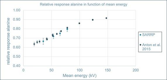
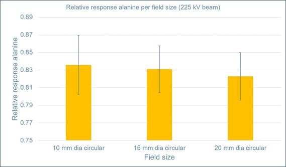
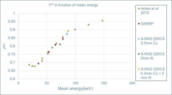
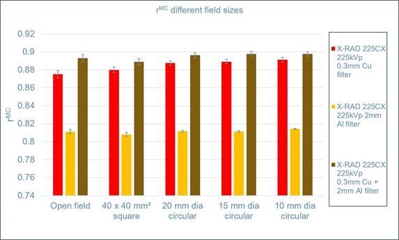
The relative response of the alanine dosimeters drops to 0.795 at photon beams with a mean energy of 78 kV and it drops further to 0.665 at photon beams with a mean energy of 35 kV. The relative response increases with 1.5% when the field size decreases to 1 x 1 cm². The simulated results are approximately 4.5 % higher than the experimental results. The intrinsic efficiency of the alanine detectors should be known to reproduce the experimental results correctly.
Experimental results
MC simulations
Type of work: Dissertation for obtaining the degree of MSc in Medical Physics at KU Leuven
Supervisor: Prof. dr. Brigitte Reniers, Hasselt University
Co-supervisor: Prof. dr. Edmond Sterpin, KU Leuven
Student: Burak Yalvac
Microdosimetry
Today, medical radiation physicists already have a wide range of equipment available to measure very accurately the amount of radiation administered to the patient (radiation dose, expressed in Gray, Gy). In addition to the absorbed dose, there is another - equally important - aspect of ionising radiation, which is the radiation quality.
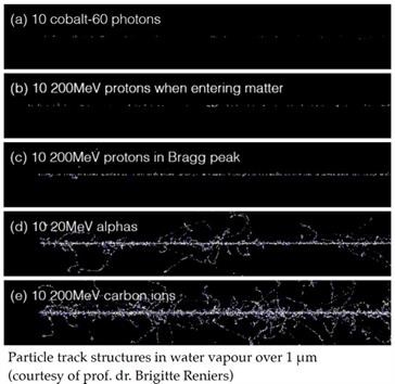
The radiation quality parameter concerns the estimation of the damage caused by radiation when crossing living tissues. As can be seen in the picture, the difference can clearly be observed between low-LET (sparsely ionising) radiation (figure a, b and c) and high-LET (densely ionising) radiation (figure d and e).
However, this radiation quality is not simply measurable in clinical conditions. Experiments can be carried out, for example, using cell cultures or live animals to evaluate radiobiological effectiveness (RBE), but the wide range of materials required for these measurements make frequent use in a (busy) hospital environment impossible.
Microdosimetry may provide a solution. Microdosimetry consists in analysing the energy depositions at the microscopic level to try to assess the effect on biological cells.
This “biologically relevant” dosimetry is clearly important in radiotherapy as soon as other types of treatment beams are considered than the megavolt photon beams used traditionally in radiotherapy (6 to 18 MV) such as in the case of the low energy photons used in brachytherapy (20 to 400 keV) or the particles used in ion beam therapy, mainly protons. It can also play a significant role to better quantify the radiobiological consequences of imaging done with photons of 30 to 120 kVp.
The research project of drs. ing. Dries Colson aims to develop a method based on the use of a physical dosimeter, a diamond based microdosimeter, that can be used for routine use in a clinical environment. The result of the physical measurements will then be related to the relative biological effectiveness (RBE) for a specific biological endpoint. In this way, the use of this microdosimeter can be used to determine the radiation quality in clinical circumstances.
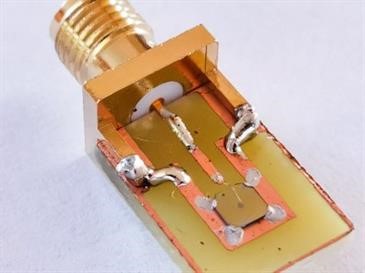
Prototype diamond microdosimeter
“Diamond detector based microdosimetry: key solution in the determination of the radiobiological effectiveness?”
https://www.uhasselt.be/project_details?pid=18125&t=en
Contact
drs. ing. Dries Colson