Meet-the-jury symposium: "From Structure to Function – Understanding Biomolecules with the Toolkit of Biophysics"
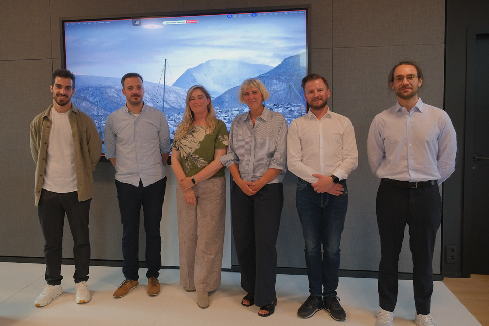
On the occasion of the PhD defenses of Pedro Silva and Tom Kache, the Dynamic Bioimaging Lab and the Advanced Optical Microscopy Centre are organizing a MiniSymposium.
Join us for three exciting lectures by experts in the biophysics field!
Practical information
Date: Friday, September 4 at 2PM.
Location: UHasselt, Refugiehuis.
Attendance: Attendance is free, but kindly confirm your presence by registering.
[registration is closed]
Programme
Joelle Goulding (Uni Nottingham): Unpicking Membrane Protein Organisation: A Fluorescence and Ligand-Directed Labelling Approach
Joelle Goulding (Uni Nottingham): Unpicking Membrane Protein Organisation: A Fluorescence and Ligand-Directed Labelling Approach
Summary
The organisation of membrane proteins on the plasma membrane can dictate drug affinity, cell signalling pathways, and subsequent cellular function. Therefore, understanding this organisation at physiologically relevant levels and within primary cell types is essential to build up our molecular pharmacology backbone to enable more effective and efficient drug development. To achieve this, I apply advanced light microscopy techniques — including fluorescence correlation spectroscopy and fluorescence lifetime imaging — alongside novel ligand-directed probes that allow us to study native receptors within key cell types. In this talk, I will present our latest work imaging adenosine receptors at endogenous expression levels
Bio
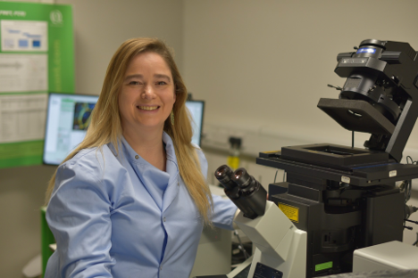
Dr Joëlle Goulding is a Senior Research Fellow in Advanced Microscopy within the Centre for Membrane Proteins and Receptors (COMPARE) at the University of Nottingham. She gained a PhD in Genetics at the University of Nottingham before moving into molecular pharmacology, developing a love of microscopy along the way. She specialises in fluorescence correlation spectroscopy but has recently introduced FLIM applications to the group's toolkit. Her current research applies advanced microscopy modalities to better understand the spatio-temporal organisation of membrane proteins, and the impact this may have on their molecular pharmacology.
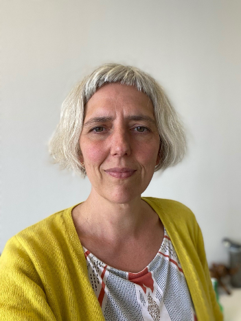
Katrien Remaut (UGhent) - From Antisense Oligonucleotide Degradation to Ocular mRNA Delivery: FCS contributions to My Academic Journey
Katrien Remaut (UGhent) - From Antisense Oligonucleotide Degradation to Ocular mRNA Delivery: FCS contributions to My Academic Journey
Summary
This presentation gives an overview on how FCS has contributed to tackle research questions related to the optimization of nucleic acid delivery using non-viral nanoparticles, with applications ranging from nucleic acid degradation, nanoparticle encapsulation, intracellular delivery and gathering new insights into the co-delivery of nucleic acids.
Bio
Prof. Dr. Katrien Remaut is an Associate Professor in the Faculty of Pharmaceutical Sciences at Ghent University, where she leads research at the Laboratory of General Biochemistry and Physical Pharmacy. Her work focuses on advanced drug and gene delivery systems, particularly lipid nanoparticles, mRNA delivery, ocular gene therapy, and nanomedicine-based treatments for cancer. She obtained her PhD in Pharmaceutical Sciences from Ghent University in 2007 and has built an internationally recognized research program on non-viral nucleic acid delivery and nanomedicine. She is also active in teaching biochemistry and contributes to the scientific community through editorial roles in leading pharmaceutical journals
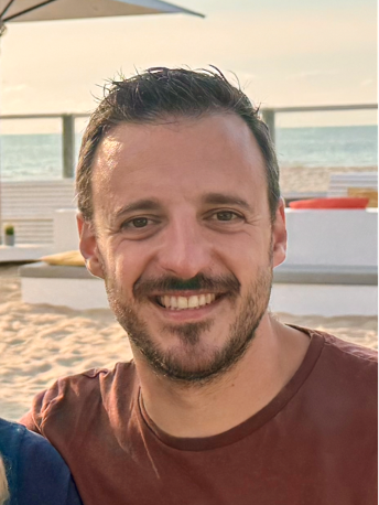
Guillaume Roussel (Rega KUL) - Mechanisms of chaperone handover: SecB and SecA dynamically tune client folding for targeted translocase delivery
Guillaume Roussel (Rega KUL) - Mechanisms of chaperone handover: SecB and SecA dynamically tune client folding for targeted translocase delivery
Summary
Bacterial preproteins synthesized in the cytoplasm must remain soluble and unfolded prior to reaching the translocase for secretion across the plasma membrane. To prevent off-pathway cytosolic aggregation while avoiding premature folding, preproteins are routed through a tightly coordinated chaperone relay, transferring from the holdase chaperone SecB to the motor ATPase SecA.
Here, we combine single-molecule Fluorescence Resonance Energy Transfer (smFRET) and Hydrogen-Deuterium Exchange Mass Spectrometry (HDX-MS) with an engineered slow-folding model client to capture conformational dynamics during SecB-to-SecA delivery. We demonstrate that SecB acts sequentially as an unfoldase to revert partially folded elements and subsequently as a holdase to preserve the unfolded state. Handover to SecA proceeds through a transient, obligate ternary complex (SecB–client–SecA), while a single-point mutation in the client facilitated the client release from SecB to SecA.
Collectively, our findings uncover how single-molecule conformational tuning and kinetic coordination ensure efficient, directional client delivery to the translocase while safeguarding against cytosolic aggregation.
Biography
Guillaume Roussel began his academic journey at the Université de Namur, where he earned his PhD in Biochemistry with a focus on designing refolding protocols for membrane proteins. Following his doctoral studies, he gained international expertise through postdoctoral positions at the Université de Montréal in Canada and the University of California, Irvine, in the USA building interests in protein dynamics and their interaction with the membrane. Currently a research associate at the Rega Institute at KU Leuven in the Laboratory of Molecular Bacteriology, his research focuses on the transport of secretory proteins across membranes and the role of chaperones in maintaining these proteins in an unfolded state; combining biochemical, biophysical and structural approaches.
Flanders BioImaging
Flanders BioImaging (FBI) is an interuniversity consortium dedicated to biomedical imaging and advanced light microscopy, that was set up to integrate, optimize, rationalize and coordinate available imaging infrastructure in Flanders, facilitating access to external users.
In 2024 the consortium fully joined EU EuroBioImaging (EuBi) project, which achieved European Research Infrastructure Consortium (ERIC) status in 2019.
FBI also coordinates with EuBI, the Flemish Research Data Network (FRDN) and the ELIXIR ERIC to develop Open and FAIR data management tools for biological and biomedical imaging in Flanders and beyond.
Contact
AOMC
Agoralaan, Building C, 3590 Diepenbeek