Lightsheet microscope launch event
The Biomedical Research Institute recently expanded its imaging portfolio with a state-of-the-art Light-Sheet Fluorescence Microscope! This full-blown platform, funded via the FWO Heavy-Scale Research Infrastructure Fund, is capable of fast tomographic imaging of living model organisms or organoids, but can also image larger cleared samples (up to 2 cm!) in toto with sub-cellular sharpness!
To introduce you to the world of light-sheet microscopy, and provide a sneak peek at what this system can mean for biomedical and clinical research, we cordially invite you to this Launch event!


Practical information
Date: Tuesday May 16, 2023 at 6:30PM
Location: UHasselt, Campus Diepenbeek, auditorium H3 (reception in Wintertuin)
Attendance: Attendance is free, but kindly confirm your presence by registering here!
Registration is now closed.

Programme
6:30PM - Welcome by prof. Jelle Hendrix, Hasselt University
6:30PM - Welcome by prof. Jelle Hendrix, Hasselt University
6:40PM - "Discovering more with the Zeiss Lightsheet 7" by Sven Terclavers, Zeiss
6:40PM - "Discovering more with the Zeiss Lightsheet 7" by Sven Terclavers, Zeiss
Summary: Lightsheet microscopy has been around for many years now, yet more and more applications are being developed. The last years, we observed an increase in cleared tissue imaging, after an initial start in life science imaging. With the introduction of lattice light sheet microscopy, the technology pushed its boundaries even further. However, light sheet imaging also comes with challenges such as sample handling, but mostly, data handling. Whereas 20 years ago, 1Tb would hold all information gathered by a small lab in one year, nowadays one single image can hold that much data. With that in mind, ZEISS is also working on several solutions to enable not only data acquisition but also data handling and processing.
Short bio: After his Biotechnology studies in Leuven, Sven Terclavers ran two imaging cores at the KULeuven/VIB for about 10 years. In 2009, he joined ZEISS as PASS Benelux – a Product & Application Sales Specialist. In this role, he was responsible to support (potential) customers with microscopy questions and help them find the right tools for their research questions. From 2014 until 2018 he worked as a PASS in the United States, first covering the South-East area, followed by three years as Embedded ZEISS Specialist at the Imaging Facility of the Harvard University in Boston. Upon my return to Europe, he accepted his current role as Head of PASS EMEA-LA.
7:20PM - "3D light-sheet imaging of large-scale optically cleared human brain and prostate tissue samples" by dr. Anna Schüth, Maastricht University
7:20PM - "3D light-sheet imaging of large-scale optically cleared human brain and prostate tissue samples" by dr. Anna Schüth, Maastricht University
Summary: The ability to image human tissue samples in 3D, with both cellular resolution and a large field of view (FOVs), can improve fundamental and clinical investigations. Dr. Anna Schueth presents a co-developed light-sheet microscopy prototype, the cleared-tissue dual view Selective Plane Illumination Microscope (ct-dSPIM), capable of fast, 3D high-resolution acquisitions, of cubic centimetre sized cleared tissue. She shows that ct-dSPIM imaging is an excellent technique to quantitatively assess entire MASH prepared large-scale human tissue samples in 3D, with considerable future clinical potential in prostate cancer.
Short bio: Dr. Anna Schueth is a postdoc at the Faculty of Neuroscience and Psychology, Maastricht University and works within the CBClab on large-scale, cleared tissue light-sheet imaging of post-mortem human brain and prostate cancer samples. As a recipient of the VENI research grant, awarded by the Dutch science organisation NWO, she has been working closely with local scientists, clinicians and international industrial partners on the co-development of a light-sheet microscope prototype and quantitative 3D visualization of both brain and prostate data. The work of Anna and her group can contribute to gaining a better understanding of 3D tissue morphology, such as prostate cancer patient tumours or human brain blood vessels.
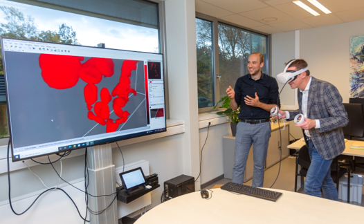
8:00PM - Reception including Virtual Reality demonstration
8:00PM - Reception including Virtual Reality demonstration
Our Zeiss Light-Sheet 7 generates large amounts of data, and is therefore equipped with a cutting-edge analysis platform. To assist users in navigating and annotating in large tomographic recordings, we provide the option to observe data in Virtual Reality. During the reception, you can have first-hand experience in such analysis. So stick around after the talks and test it for yourself!


Flanders BioImaging
Flanders BioImaging (FBI) is an interuniversity consortium dedicated to biomedical imaging and advanced light microscopy, that was set up to integrate, optimize, rationalize and coordinate available imaging infrastructure in Flanders, facilitating access to external users.
In 2022 the consortium was successfully evaluated as a candidate note by the EU EuroBioImaging (EuBi) project, which achieved European Research Infrastructure Consortium (ERIC) status in 2019.
FBI also coordinates with EuBI, the Flemish Research Data Network (FRDN) and the ELIXIR ERIC to develop Open and FAIR data management tools for biological and biomedical imaging in Flanders and beyond.
Contact
AOMC
Agoralaan, Building C, 3590 Diepenbeek